As humans, we are susceptible to several sicknesses. When we fall sick, we need to get diagnosed and get treated.
Sometimes, the name of some medical operations may sound so grave and intimidating: For instance, there is “Bronchoscopy”. When you hear the word; it may sound a little too much. However, it is simply a treatment or diagnostic procedure that allows a healthcare provider to look inside their patient’s lungs and airways.

Equipment like a BodyVision Imaging is often used for the procedure. The process is also not as scary as it sounds. In case you need to get this process done, then you don’t have to be scared. To help ease your mind, we will be discussing how this procedure is done. This way, you will know just what to expect.
Bronchoscopy Procedure
The whole process would typically last somewhere between 30 minutes and some hours. It is an outpatient process. The following are what happens during bronchoscopy:
- The patient will be asked to lie on either a table or a bed, and their head would also be slightly raised up.
- An IV will be inserted into the patient’s arm and a sedative will be passed through it to help them relax. Some persons often wish to sleep throughout the process. General anesthesia is often used for rigid bronchoscopy. You can speak to your healthcare provider so a conclusion will be made about whether or not you need general anesthesia or if it even suits your needs.
- A spray is applied to the patient’s throat and nose or mouth to numb them.
- Now that the patient is sedated and the areas for the process are numb, the bronchoscope (the equipment for the process) is inserted either through the mouth or nose. The bronchoscope will go down the windpipe and down to the lungs.
- Once the healthcare provider takes a look at the lungs and airways, the bronchoscope will be gently removed.
The process is complete at this point. See, we told you it wasn’t so difficult. After the process is complete, the patient’s condition will be monitored by the medical team until they wake up.
After the Procedure – What Next
Because it’s an outpatient process, you don’t have to sleep overnight in the health facility. You can read this to learn more about outpatient surgeries and procedures. You can even leave the healthcare facility some hours after the process.
You will be monitored by the medical team to make sure you are swallowing and breathing properly. The numbness you feel in the throat that is caused by the spray may last for some hours before it completely goes away.
You may experience cough, hoarseness in the throat, and sore throat for a day after the process. You will also be told when you will get the results; follow-up dates will be given as well. The result should typically be ready in some days.

Bronchoscope
The bronchoscope is the equipment used for this medical process. Also, an image retrieval device that has a light-generating device would be connected to the bronchoscope with a cord.
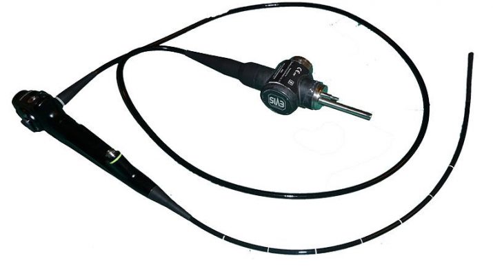
A flexible bronchoscope typically has 3 parts.
Parts of a Flexible Bronchoscope
The following are the parts of a flexible bronchoscope:
- Control Handle: The operator has to hold the bronchoscope’s control handle. This part has a lever that is used to extend or flex the bronchoscope’s distal tip. The handle also has an opening that other instruments like brushes, forceps, and so on will be inserted into. A suction port is also on this part.
- Distal Tip (Working Tip): This is what will be inserted into your airway. It has a light source, the working channel opening, and image retrieval (camera).
- Flexible Shaft: This part has cables that are enclosed by a sheath and it allows the extension and flexion of the bronchoscope’s distal tip. The handle’s lever is moved to do this. Imaging cables, the working channel for aspirating airway contents and passing in other instruments, and lighting cables are also on this part.
Light Source
As we have said, a light source is often connected to the equipment. This is used to transmit white light. The light’s brightness, color, and intensity can be adjusted.
Image Processor
The airway would be illuminated by the light source, this would make imaging possible. The image gotten will be transmitted either via digitized video transmission or fiberoptic transmission.
The original image transmission method is the fiberoptic transmission. However, the newer digitized video transmission is now available. Visit https://www.techtarget.com/searchnetworking/definition/fiber-optics-optical-fiber to learn more about fiber optics.
The new method uses a small charge-coupled tool that is fixed at the bronchoscope’s working tip. The images that are gotten via this method are magnified, shaper, and of higher resolution.
Conclusion
We believe you now know what to expect during a bronchoscopy procedure. We hope this article has helped put your mind at ease and prepare you for the process.